Nikon announced the best photos of 2018 made using microphotography
Nikon has been hosting the Nikon Small World Photomicrography Competition for 44 years in a row. The competition was established in 1974, but so far its participants continue to amaze with fantastic pictures of their observations made through the lens of a light microscope. Despite the fact that there are many scientists among the contestants, anyone can participate in Nikon Small World. Organizers do not limit the subject of images; the authors are also completely free to choose the technique of microphotography.
The winners of 2018 have already been announced, and now we will introduce you to their impressive works.
The first place went to the photographer Yussef Al-Habshi from the UAE. In his photograph of the eye of the beetle Metapocyrtus subquadrulifer, magnified 20 times, he applied the technique of reflected light. But that's not all: the photo is a compilation of 128 microphotographs.

In second place is Rochelio Moreno from Panama and his snapshot of suss ferns. Sorus is a part of a plant that produces and stores spores. It is noteworthy that with the help of ultraviolet, the author was able to distinguish spore-bearing organs in different colors, thereby capturing various levels of spore maturity.

Third place went to American Saulius Gugis. In his photo we can see the Spittlebug cicada-pennant doll in her bubble house, which she builds to protect herself from predators and cold weather.

Kan Tunser, Turkey. Peacock feather, enlarged 5 times.

Tessa Montague, USA. Parasteatoda tepidariorum spider embryo, enlarged 20 times.

Hanen Habu, France. Foveola primate. Or, more simply, the central region of the retina, enlarged by 40 times.

Norm Barker, USA. A person’s tear, increased by 5 times.

Pia Scanlon, Australia. Mango weevil.

Haris Antonopoulos, Greece. Security hologram magnified 10 times.

Chaba Pinter, Hungary. Stems with pollen, increased 3 times.

Nilai Taneja and Dylan Burnett, USA. Human fibroblast at the time of cell division, increased by 60 times.

Luciano Andres Rihino, Argentina. Butterfly wing enlarged 20 times.

Charles Krebs, USA. Balanus glandula, increased by 5 times.

Andrew Moore and Erica Holzbauer, USA. African green monkey cell (COS-7), increased 100 times.

Antoine Frank, Saint Pierre, Seychelles. The parasitic tick Varroa destructor on the body of a honey bee.

Amanda Phillips-Isaguerre, USA. Vascular network of the mouse, increased by 10 times.

Caleb Dawson, Australia. Breast tissue during lactation. An increase of 63 times.

Justin Zoll, USA. Amino acid crystals of L-glutamine and beta-alanine, increased 4 times.

Pierre Questionnaire, France. The Asian hornet Vespa velutina, on the sting of which a drop of poison is visible.

Nicholas Cuenca and Isabelle Ortuno Lisaran, Spain. The retina of a person’s eye, increased by 40 times.

Watch the video: A Must Have Nikon Macro Lens. Nikon 105mm Micro Review by Georges Cameras (May 2024).

-
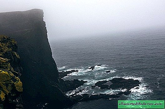
Iceland: on the verge of death
We got to Western Fjords, where, according to the guide, there are just millions of dead ends. However, the reality was completely different ... There were no dead ends! It turned out that the dead end season ended 5 days ago and they had all left. But I was lucky to see the heels of the last lagging dead end disappearing in the fog. ... -
-
-